Mock ARDMS AE-Adult-Echocardiography Exam & AE-Adult-Echocardiography Reliable Dump
Wiki Article
P.S. Free & New AE-Adult-Echocardiography dumps are available on Google Drive shared by Exam4Docs: https://drive.google.com/open?id=1h4soc-ceY8voNFiXFg39G702RxdvboDu
Students are worried about whether the AE-Adult-Echocardiography practice materials they have purchased can help them pass the exam and obtain a certificate. They often encounter situations in which the materials do not match the contents of the exam that make them waste a lot of time and effort. But with AE-Adult-Echocardiography exam dump, you do not need to worry about similar problems. Because our study material is prepared strictly according to the exam outline by industry experts, whose purpose is to help students pass the exam smoothly. As the authoritative provider of AE-Adult-Echocardiography Test Guide, we always pursue high passing rates compared with our peers to gain more attention from potential customers.
ARDMS AE-Adult-Echocardiography Exam Syllabus Topics:
| Topic | Details |
|---|---|
| Topic 1 |
|
| Topic 2 |
|
| Topic 3 |
|
| Topic 4 |
|
| Topic 5 |
|
>> Mock ARDMS AE-Adult-Echocardiography Exam <<
AE-Adult-Echocardiography Reliable Dump, AE-Adult-Echocardiography Best Practice
Without no doubt that accuracy of information is of important for a AE-Adult-Echocardiography study material. It can be said exactly that the precision and accuracy of our Exam4Docs’s AE-Adult-Echocardiography study materials are beyond question. All questions and answers have passed the test of time and are approved by experienced professionals who recommend them as the easiest route to certification testing. Every customer who has used our AE-Adult-Echocardiography Study Materials consider this to be a material that changes their life a lot, so they recommend it as the easiest way to pass the certification test. Our AE-Adult-Echocardiography study materials are constantly updated by our experts and improved according to the changing standards of the actual examination standards. We can guarantee that the information on our questions is absolutely true and valid.
ARDMS AE Adult Echocardiography Examination Sample Questions (Q26-Q31):
NEW QUESTION # 26
Which method is appropriate for measuring the left atrial diameter in parasternal long axis?
- A. Inner edge to inner edge, parallel to the aortic root, at end-diastole
- B. Inner edge to inner edge, perpendicular to the aortic root, at end-diastole
- C. Inner edge to inner edge, perpendicular to the aortic root, at end-systole
- D. Outer edge to outer edge, perpendicular to the aortic root, at end-systole
Answer: B
Explanation:
Comprehensive and Detailed Explanation From Exact Extract:
The recommended method to measure left atrial diameter in the parasternal long axis view is the inner edge to inner edge technique, perpendicular to the aortic root, measured at end-diastole. This approach provides the most reproducible and standardized measurement.
Measurement parallel to the aortic root or at end-systole is less accurate. Outer edge measurements overestimate size.
ASE chamber quantification guidelines specify this method for standardization and reproducibility in adult echocardiography practice#12:ASE Chamber Quantification Guidelinesp.90-95##16:Textbook of Clinical Echocardiography, 6ep.120-125#.
NEW QUESTION # 27
Which finding is most commonly associated with Ebstein anomaly?
- A. Atrial septal defect
- B. Tricuspid stenosis
- C. Ventricular septal defect
- D. Pulmonary stenosis
Answer: A
Explanation:
Comprehensive and Detailed Explanation From Exact Extract:
Ebstein anomaly is a congenital malformation characterized by apical displacement of the tricuspid valve leaflets, leading to atrialization of the right ventricle and severe tricuspid regurgitation. The most common associated defect is an atrial septal defect (ASD), particularly a secundum type or patent foramen ovale, resulting in right-to-left shunting and cyanosis.
Ventricular septal defect and pulmonary stenosis are less commonly associated. Tricuspid stenosis is not typical; the tricuspid valve is usually regurgitant rather than stenotic.
This association is well described in congenital heart disease and echocardiography textbooks and ASE guidelines#16:Textbook of Clinical Echocardiography, 6ep.570-575##12:ASE Adult Congenital Guidelinesp.400-405#.
NEW QUESTION # 28
Which echocardiography assessment requires mitral inflow pulsed wave, pulmonary venous pulsed wave, and tissue Doppler of the mitral annulus?
- A. Mitral regurgitation severity
- B. Left ventricular diastolic function
- C. Left ventricular systolic function
- D. Myocardial performance index
Answer: B
Explanation:
Assessment of left ventricular diastolic function by echocardiography involves evaluating mitral inflow velocities with pulsed wave Doppler (E and A waves), pulmonary venous flow patterns (systolic and diastolic waves), and tissue Doppler imaging of the mitral annulus to measure early diastolic (e') velocities.
This combination allows differentiation of normal versus abnormal relaxation, elevated filling pressures, and grading of diastolic dysfunction. The myocardial performance index evaluates global ventricular function but does not specifically require these Doppler measures. Systolic function is assessed mainly by ejection fraction and wall motion. Mitral regurgitation severity uses color Doppler and vena contracta measurements.
This multiparameter diastolic function evaluation is outlined in the "Textbook of Clinical Echocardiography,
6e", Chapter on Diastolic Function Assessment#20:210-220Textbook of Clinical Echocardiography#.
NEW QUESTION # 29
What is indicated by the arrow on this video clip?
- A. Chiari network
- B. Eustachian valve
- C. False tendon
- D. Moderator band
Answer: D
Explanation:
The structure indicated by the arrow in the right ventricle is the moderator band. The moderator band is a muscular band of tissue that crosses the right ventricular cavity from the interventricular septum to the anterior papillary muscle. It contains part of the right bundle branch of the conduction system and is a normal anatomical structure identifiable on echocardiography.
False tendons are fibrous or muscular strands within the left ventricle, not the right. The Chiari network is a mobile, net-like structure in the right atrium near the inferior vena cava and atrial septum. The Eustachian valve is a crescent-shaped ridge at the entrance of the inferior vena cava into the right atrium.
The moderator band is important to recognize to avoid misinterpretation as a pathological mass or thrombus.
This is detailed in the "Textbook of Clinical Echocardiography, 6e", Chapter on Right Ventricular Anatomy and Echocardiographic Landmarks#20:150-155Textbook of Clinical Echocardiography#.
NEW QUESTION # 30
Which patient positioning is best for obtaining the waveform seen in this image obtained by a non-imaging transducer?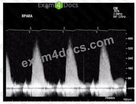
- A. Laying on left side
- B. Laying on stomach with left arm raised
- C. Laying on back with chin down
- D. Laying on right side
Answer: D
Explanation:
Comprehensive and Detailed Explanation From Exact Extract:
The image shows a Doppler waveform of the right pulmonary artery (RPARA) flow obtained using a non- imaging (pedoff) continuous wave Doppler transducer. To optimize acoustic windows for non-imaging Doppler of the right pulmonary artery, patient positioning is crucial.
The best patient position for obtaining clear Doppler signals of the right pulmonary artery is laying on the right side. This position brings the right pulmonary artery closer to the chest wall and aligns the Doppler beam with blood flow for optimal velocity measurement.
Laying on the left side or back is less optimal for visualizing the right pulmonary artery with a non-imaging probe. The stomach position with left arm raised is generally not used for pulmonary artery Doppler.
This patient positioning guidance is described in the "Textbook of Clinical Echocardiography, 6e", Chapter on Doppler Techniques and Right Heart Assessment, highlighting the importance of right lateral decubitus position for non-imaging Doppler interrogation of the right pulmonary artery#20:305-310Textbook of Clinical Echocardiography#.
NEW QUESTION # 31
......
As the most popular AE-Adult-Echocardiography exam questions in the field, the passing rate of our AE-Adult-Echocardiography learning questions has up to 98 to 100 percent. And our AE-Adult-Echocardiography preparation materials have three versions to satisfy different taste and preference: PDF version, Soft version and APP version. The three versions of AE-Adult-Echocardiography training prep have the same questions, only the displays are different. You can buy according to your interest. In addition, AE-Adult-Echocardiography test engine is indispensable helps for your success.
AE-Adult-Echocardiography Reliable Dump: https://www.exam4docs.com/AE-Adult-Echocardiography-study-questions.html
- Get the ARDMS AE-Adult-Echocardiography Certification within the Target Period ???? Open “ www.exam4labs.com ” and search for ▛ AE-Adult-Echocardiography ▟ to download exam materials for free ????AE-Adult-Echocardiography Valid Vce
- Realistic ARDMS Mock AE-Adult-Echocardiography Exam ???? The page for free download of ( AE-Adult-Echocardiography ) on ▷ www.pdfvce.com ◁ will open immediately ????AE-Adult-Echocardiography Valid Vce
- Pass AE-Adult-Echocardiography Guarantee ???? AE-Adult-Echocardiography Download ???? AE-Adult-Echocardiography Latest Exam Question ???? Search for ☀ AE-Adult-Echocardiography ️☀️ and obtain a free download on [ www.vce4dumps.com ] ????AE-Adult-Echocardiography Valid Dumps Questions
- Exam AE-Adult-Echocardiography Tips ⚔ AE-Adult-Echocardiography Valid Study Guide ???? AE-Adult-Echocardiography Download ✋ Simply search for 《 AE-Adult-Echocardiography 》 for free download on “ www.pdfvce.com ” ????AE-Adult-Echocardiography Valid Dumps Questions
- AE-Adult-Echocardiography Exam Pass4sure ???? AE-Adult-Echocardiography Reliable Practice Questions ???? AE-Adult-Echocardiography Top Exam Dumps ???? Download ▛ AE-Adult-Echocardiography ▟ for free by simply entering [ www.prepawayete.com ] website ????AE-Adult-Echocardiography Valid Vce
- 100% Pass 2026 AE-Adult-Echocardiography: High Pass-Rate Mock AE Adult Echocardiography Examination Exam ???? Search for ➠ AE-Adult-Echocardiography ???? and obtain a free download on 「 www.pdfvce.com 」 ⌛AE-Adult-Echocardiography Latest Test Preparation
- Mock AE-Adult-Echocardiography Exam ???? Valid AE-Adult-Echocardiography Exam Testking ???? New AE-Adult-Echocardiography Test Simulator ???? Search on { www.prepawayexam.com } for 【 AE-Adult-Echocardiography 】 to obtain exam materials for free download ????AE-Adult-Echocardiography Latest Test Preparation
- AE-Adult-Echocardiography Latest Exam Question ???? AE-Adult-Echocardiography Download ???? Free AE-Adult-Echocardiography Download ???? Simply search for ☀ AE-Adult-Echocardiography ️☀️ for free download on ⮆ www.pdfvce.com ⮄ ????AE-Adult-Echocardiography Top Exam Dumps
- AE-Adult-Echocardiography Reliable Practice Questions ???? Free AE-Adult-Echocardiography Download ???? AE-Adult-Echocardiography Latest Exam Preparation ???? Immediately open [ www.testkingpass.com ] and search for ➥ AE-Adult-Echocardiography ???? to obtain a free download ????AE-Adult-Echocardiography Latest Exam Question
- Quiz Useful AE-Adult-Echocardiography - Mock AE Adult Echocardiography Examination Exam ???? Immediately open [ www.pdfvce.com ] and search for ▛ AE-Adult-Echocardiography ▟ to obtain a free download ????AE-Adult-Echocardiography Latest Exam Preparation
- AE-Adult-Echocardiography Valid Dumps Questions ???? Mock AE-Adult-Echocardiography Exam ???? AE-Adult-Echocardiography Valid Study Guide ???? Easily obtain ➡ AE-Adult-Echocardiography ️⬅️ for free download through ☀ www.prepawaypdf.com ️☀️ ????Mock AE-Adult-Echocardiography Exam
- laralfff102992.blog-a-story.com, teganoftl515745.blogdal.com, bookmarksfocus.com, social-galaxy.com, agnesnpyn864091.bloggadores.com, shaunauewy944851.angelinsblog.com, nitizsharma.com, www.stes.tyc.edu.tw, chiaraqhay975961.goabroadblog.com, robertfhai936310.ambien-blog.com, Disposable vapes
P.S. Free & New AE-Adult-Echocardiography dumps are available on Google Drive shared by Exam4Docs: https://drive.google.com/open?id=1h4soc-ceY8voNFiXFg39G702RxdvboDu
Report this wiki page